
Luckily, we are highly skilled in treating a variety of conditions and issues. Whether you have slight discomfort or severe pain, we can provide you with care that allows you to live the lifestyle you want!
These are just some of the conditions, treatments, and services you will find at our podiatric office. Just ask us if you have any questions!
- Achilles Tendon Rupture
- Ankle Fractures
- Ankle Sprains
- Arthritic Foot/Ankle Problems
- Ball of Toe Pain
- Bunions
- Charcot Foot
- Chronic Swelling
- Cysts of the Foot or Leg
- Hammertoes
- Heel Pain
- Lapiplasty
- Neuromas
- Peripheral Arterial Disease
- Platelet Rich Plasma Injection
- Posterior Tibial Tendon Dysfunction
- Preventive Foot Care
- Shin Splints
- Athlete’s Foot
- Corns
- Diabetic Foot Care & Diabetic Wounds
- Ingrown Toenails
- Plantar Warts
- Porokeratosis
- Sports Injuries
- Nonhealing Fractures

Achilles Tendon Rupture
Rupture of the Achilles tendon, or large tendon in the back of the lower leg, can be a potentially debilitating injury. This tendon attaches the muscles in the back of the leg to the back of the heel bone, allowing us to push our weight off of the balls of the toes.
As we age, the elasticity of our tissues, including the Achilles tendon, declines with age. One particular tissue, Elastin, tends to try out with age, making us more prone to rupturing the Achilles tendon with each passing year.
Achilles tendon injuries often occur in people over the age of 30 years old. Surgical repair of the damaged tendon is usually recommended, to maintain function of the tendon. Neglecting an Achilles tendon injury can significantly reduce the success rate of surgical repair, and may potentially lead to debilitating problems, pain, and difficulty walking.
If you sustain an acute injury to your Achilles tendon, believe you may have injured your Achilles tendon, or simply have pain in your Achilles tendon, give us a call at (270) 684-5252.
Early detection and treatment is the primary way to avoid further injury and problem. If left untreated, a simple tendinitis of the tendon can progress to a full rupture, requiring a significant period of time for adequate healing.

Ankle Fractures
An ankle fracture can be a significantly traumatic event, causing significant pain and potential morbidity. Often the result of an acute injury, ankle fractures sometimes require surgery to stabilize and reapproximate the broken bones.
If left undiagnosed or untreated, this can result in significant deformity and premature arthritis in the joint, leading to significant debilitating pain in the ankle.
If you have sustained an ankle injury, or believe you may have fractured a bone, such as the ankle, please give us a call at (270) 684-5252.
We would be happy to see you during the week, as well as on the week-ends.
We provide on-site digital x-rays, and are experienced and knowledgeable and treating all aspects of foot and ankle trauma, including fractures of the foot and ankle.
Ankle Sprains
Ankle sprains are one of the most acute injuries seen in our office. A sprain is a tear or stretch of a ligament of a joint. A ligament attaches a bone to bone, and functions to provide stability of a joint.
Ankles have ligaments on all sides of the joint. Due to the shape of the ankle bones, the ankle is most vulnerable to sprains by turning the ankle “inward” with the sole of the foot facing the opposite foot. This creates a significant amount of tension on the ligaments on the outside of the ankle. This are of the ankle is most prone to sprain as a result of these factors, and is the area most likely to be painful following an ankle injury.
The most basic treatment options for a sprain include RICE (rest, ice, compression, elevation). Anti-inflammatory medications, such as ibuprofen or Aleve can also be helpful. If you have a severe sprain and are unable to walk, it is always advised to have x-rays taken to ensure one or more bones is not fractured (broken). With more severe sprains, extended immobilization in a walking boot and possibly crutches may be needed.
If you have experienced a foot or ankle sprain, and are concerned about your condition, give us a call at (270) 684-5252.

Arthritic Foot/Ankle Problems
A joint can experience significant wear and tear over the years, and may eventually wear out. Where two bones meet, a soft, slick, cushioning, pliable tissue called cartilage helps the bones glide against one another in a painless, efficient manner. Several different factors can result in the damage or loss of the cartilage, causing the bones to rub together. These factors include history of trauma, other disease such as rheumatoid arthritis, genetics, or poor mechanics. Depending upon the severity of the damage to the cartilage, pain, swelling, and possible limitation of motion of the joint results.
Depending upon where the joint is located, treatment options can vary. For early stage arthritis of the foot or ankle, anti-inflammatory medication, rest, ice, use of more supportive shoes, arch supports, steroid injection, physical therapy, and mobility exercises may offer relief. In more advanced cases of arthritis, surgery is an option. Surgery options vary widely by the particular joint involved, the severity of arthritis, age and overall health of the patient, as well as other factors.
Options can include surgery to improve the mechanics of the joint, preserving the joint, allowing more fluid motion, reducing pain, and improved function. Other cases may require fusion of the joint, where the remaining joint tissue is removed and hardware (screws and/or plates) are used to cause the two bones to grow together, removing the pain of the arthritic joint. Finally, joint replacement is an option in particular joints, including the ball of the great toe and ankle joint.
If you suffer from arthritic foot or ankle pain, or what you believe may be arthritis, give us a call at (270) 684-5252. Treatment options are available!

Ball of Toe Pain
One of the more common conditions treated in our office, is that of pain in the base of the 2nd and/or 3rd toes, caused by strain and pressure in this area with walking. This is often due to a length discrepancy of the 2nd metatarsal being slightly longer than the 1st and 3rd metatarsals. During the toe-off phase of gait, we push off the balls of our toes for propulsion. When the 2nd metatarsal is longer than the others, an increase in pressure is placed on the bottom of this area often causing pain.
Factors that can contribute to this development are excessive walking barefoot, wearing high-heeled shoes, flip-flops, sandals, house slippers, bunions, and hammer toes. Bunions can be associated with this deformity due to the foot’s inability to stabilize the medial column against the ground. When this occurs, the body weight is transferred to the adjacent, more stable metatarsal, leading to the pain below the 2nd metatarsal head. Also, contracture of the toes can lead to this type of pain, as the curling of the toes causes the base of the proximal phalanx to ride on the dorsal aspect of the metatarsal head, causing the foot to feel like the metatarsal head is protruding out the bottom of the foot.
Treatment is focused on reduction of the inflammation by way of arch supports, avoidance of barefeet, flip flops, sandals, and shoes with a heel, as well as ice applied below ball of toes x 20-25 minutes 4-5 times a day. Anti-inflammatory medication can also help.
In a few number of cases, conservative measures don’t result in pain resolution. Some of these patients may be reluctant to pursue heel spur surgery or have continued pain following surgery. For these patients we offer a comprehensive treatment plan to offer hope. These modalities are not covered by insurance, but could very likely result in resolution of pain. By way of a combination of up to 10 MLS laser treatments, focused on resolving the inflammation and scar tissue in the area, along with Extra Corporeal Shockwave Treatments, and possibly, Platelet Rich Plasma Injection, we are confident we can resolve some, if not all of your chronic heel pain. The cost of the comprehensive plan can vary from $2,000 – $3,500 for a comprehensive plan spanning 5-12 months to alleviate resistant heel pain cases.
If you suffer from foot pain, give us a call at (270) 684-5252. Treatment options are available!
Bunions
A bunion is a bony bump that forms on the joint at the base of your big toe. They can develop from an inherited structural defect, excess stress on your foot, or can result from an existing medical condition.
For the most part, bunions require no medical treatment. However, if you are experiencing one or more of the following, a podiatrist can help alleviate your symptoms.
A bunion (hallux valgus) is a progressive deviation of the big toe towards the second toe and development of a knot on the ball of the big toe. This inherited deformity is very common and effects women more than men. Bunons occur can occur at any stage of life, but are more common in those over 50 years of age.
The cause is due to an imbalance of the tendons attached to the base of the big toe. Like reigns on a horse, if one reign is pulled tighter than the other, the horse turns. Similarly, the inherited discrepancy of tension between the two sides of the great toe gradually cause the toe to drift towards the second toe.
Development of a bunion is primarily genetic and prevention is aimed at reducing the forces causing the big toe to lean towards the second; such as avoidance of narrow toed shoes.
Treatment options involve accommodative padding, wide shoes to reduce the pressure against the ball of the big toe, or surgery.
Surgical treatment varies widely by physician, as does the level of mobility allowed after surgery. Also, the actual techniques used to correct the bunion can also vary by provider and can alter the amount of mobility permitted in the post-operative period.
Most bunion surgeries are performed as an outpatient, where the patient arrives to the hospital or surgical center, has the procedure, and goes home all in the same day. The surgery is also frequently performed under IV sedation, also called “twilight anesthesia”. This involves insertion of an intravenous (IV) line into the arm or hand. Medications to make the patient dose off to sleep will then be administered allowing pain free injection of local anesthetic into the surgical site. The patient does not feel the injections, does not inhale gas, have a tube down their throat, nor do they experience the nausea and vomiting often associated with a general anesthetic.
The surgery takes an average of 30-60 minutes. Depending upon the severity of the bunion, the amount of activity permitted after the surgery may range from walking in a removable walking boot to a below knee cast, complete nonweight bearing, and use of crutches.
Typically, bunion surgery involves use of a walking boot, where the patient is allowed to walk on a limited basis beginning the same day of surgery. This involves walking to the restroom, kitchen, car, etc. Prolonged walking or standing is not advised. Primary bone healing occurs in 4-6 weeks, and may extend to 8 weeks for smokers. The patient is allowed to return to a large tennis shoe at about four weeks, pending everything looks good both clinically and on x-ray. The foot will continue to be wrapped with a compressive wrap in the shoe for up to two more months to minimize swelling.
During the first four weeks after surgery, the bones are healing, but not fully healed. Like glue partially dried, the bones that were corrected are fragile. If the foot is not sufficiently splinted or stabilized, and should the foot be jarred or a toe be stubbed, the surgical site can become dislodged and positioned out of alignment, resulting in a return of the deformity, continued pain, or a return to the operating room for repairs.
Virtually all insurance plans cover elective bunion surgery and verification of benefits for this surgery is performed prior to your surgery.

Charcot Foot
Charcot neuroarthropathy is a potentially debilitating condition associated with diabetes, causing the bones of the foot to shatter. The reason why Charcot develops is not completely known. It is much more common in neuropathic (loss of sensation in the feet) diabetic patients who are insulin-dependent and has been for 10 or more years.
When Charcot develops, there is an influx of blood flow to one certain area of the foot or ankle, which eventually leads to the calcium and other minerals of the bone being washed away. The acute phase of Charcot last 1-2 weeks, and is exhibited by significant swelling, redness, and heat produced by either the entire foot or a particular region of the foot or ankle.
During the acute phase of Charcot, if weightbearing continues, the joints and bones of this region of the foot can become very brittle and fracture into multiple shards of bone, resulting in severe deformity of the foot. If immediate nonweightbearing is assumed immediately when the foot or ankle is noted to be red, hot, and swollen, the destruction and deformity often associated with Charcot can be avoided.
Typically during the acute phase of Charcot, nonweightbearing is required for up to 4 weeks. If x-rays appear that the acute phase is pending, then the weightbearing can be resumed, with some extra compression such as with compression hose to minimize swelling and some protection in a walking boot or walking cast.
It is the deformities associated with Charcot that often lead to other chronic diabetic foot wounds, which can lead to other significant problems including the need for amputation.
Maintaining regular visits with a physician to monitor glucose levels, and regular visits with a foot specialist can help prevent the deformities and Associated problems of diabetes.
If you suffer from foot pain, give us a call at (270) 684-5252. Treatment options are available!

Chronic Swelling
One of the most common yet misdiagnosed conditions we see at the Bluegrass Foot Center is superficial thrombophlebitis (thrombo flibytis). Chronic swelling of the legs is very common, and occurs for a number of reasons. Chronic high blood pressure, obesity, liver failure, venous insufficiency, history of numerous pregnancies, pelvic or groin surgery, radiation of pelvis or groin region, venous reflux, and inactivity are all potential reasons why one may develop chronic swelling of the legs. All of these conditions cause a reduced ability of the veins of the legs to return blood to the body. When this occurs, fluid builds up in the legs, resulting in swelling of the legs.
Our skin is like a balloon. If you blow up a balloon too much it pops. Similarly, as the legs swell, the skin gets irritated and inflamed, causing the skin to become red, hot, and tender to touch. If left untreated, the skin can develop sores, or venous stasis ulcerations.
Frequently, other health providers view the redness as an infectious process, called cellulitis. The patient is commonly admitted to the hospital, placed on IV antibiotics and in a hospital bed. This treatment often resolves much of the redness and swelling, not as a result of the antibiotics, yet due to elevation of the legs, resulting in the reduction of the redness and swelling. In fact, in most cases, infection was never present.
Use of below the knee compression hose can effectively control the chronic swelling of the legs, reducing the recurrence of phlebitis (inflammation of the vessels of the skin). Compression hose are sold in the office,and are fit based on size of the legs. In addition, a lymphedema pump can also be of assistance. This involves wrapping the legs with inflatable bladders, with a machine elevating the bladders to squeeze on the legs at set time intervals. Finally, when the patient is either unable to tolerate or unable to apply compression hose on a daily basis, once weekly application of wraps, called Unnaboots, can help. These comfortable wraps are left clean, dry, and intact for 7 days, until the following week, at which time new wraps are applied.
The weekly application of wraps is indefinite. Unfortunately, any external compression treatment for treatment of venous insufficiency or phlebitis is transient, and needs to be employed indefinitely. Chronic swelling and repeat episodes of superficial thrombophlebitis can result in scarring of the skin, called post phlebitic syndrome. This condition can result in chronic wound formation of the skin, and may result in having to apply unnaboots to the legs on a once weekly basis indefinitely.
If you suffer from chronic swelling of the legs or frequent redness and tenderness of the lower legs, give us a call at (270) 684-5252. Treatment options are available!

Cysts of the Foot or Leg
A ganglion cyst is a collection of fluid around either a joint or tendon. A tendon, which attaches a muscle to bone, has an outer covering called a tendon sheath. Between the tendon and the tendon sheath is fluid which acts to lubricate the tendon and reduce friction as the tendon moves within the sheath.
Likewise, a joint, where two bones touch one another, has a balloon (joint capsule) around the outside of these bones. Within this balloon is fluid (synovial fluid) which also acts to lubricate and ease motion of the joint.
A traumatic event, either recognized or unnoticed, can cause the tendon sheath or joint capsule to herniate or balloon outward, called a ganglion cyst. This herniated area fills with fluid, frequently causing an enlarged, painful mass. Frequently, the mass will increase and decrease in size, varied by the amount of fluid in the cyst. Ganglion cysts can occur around any tendon or joint. Treatment options include either injecting the cyst with a steriod to reduce its size, draining the fluid of the cyst by insertion of a needle (which usually results in a quick return of the cyst), or surgical excision.
If you suffer from foot pain, give us a call at (270) 684-5252. Treatment options are available!

Hammertoes
A hammer toe is defined as a contracture of the toe. Caused by an imbalance of the tendons on top and bottom of the toes, a hammer toe is initially flexible, where it can manually be straightened with a finger. With time, the flexibility of the toe with be reduced, leaving the toe rigidly contracted. A rigid hammer toe is not only painful to touch over the knuckle, but it is also painful with attempted straightening of the toe.
Most commonly, hammer toes cause pain with shoes as the top of the shoe rubs on the top of the toes, causing redness, callouses, and occasionally sores to develop. Treatment is aimed at reduction of symptoms by either padding the irritated area with over the counter (non medicated) corn pads to prevent the shoes from rubbing on the area, wearing larger shoes with more room in the toe area, or surgically reducing the contracted toe.
If you suffer from foot pain, give us a call at (270) 684-5252. Treatment options are available!

Heel Pain
One of the most common complaints in our office is that of heel pain. In excess of ninety percent of heel pain complaints are diagnosed as having inflammation of the plantar fascia (a ligament on the bottom of the foot). Commonly referred to as a heel spur or stone bruise, plantar fasciitis (plantar fashitis) can be a severely debilitating and painful condition.
Frequently described as a deep burning ache, the pain is most commonly experienced early in the morning when first placing the feet on the ground, and also following a short period of rest, for example, when getting up to walk after watching television for an hour or two.
What causes the pain is any one or combination of things. A change in shoe gear, an increase in physical activity, lack of adequate support in shoes, frequently walking barefoot, an increase in weight, or simply a naturally flexible foot type all contribute to the development of plantar fasciitis.
In a few number of cases, conservative measures don’t result in pain resolution. Some of these patients may be reluctant to pursue heel spur surgery or have continued pain following surgery. For these patients we offer a comprehensive treatment plan to offer hope. These modalities are not covered by insurance, but could very likely result in resolution of pain. By way of a combination of up to 10 MLS laser treatments, focused on resolving the inflammation and scar tissue in the area, along with Extra Corporeal Shockwave Treatments, and possibly, Platelet Rich Plasma Injection, we are confident we can resolve some, if not all of your chronic heel pain. The cost of the comprehensive plan can vary from $2,000 – $3,500 for a comprehensive plan spanning 5-12 months to alleviate resistant heel pain cases.
Treatment is directed at minimizing the pull and tension of the plantar fascia with standing and walking. Treatment must involve mechanical support of the arch to reduce the tension and strain of the plantar fascia, AND reduction of the inflammation of the fascia attachment at the heel bone. Simply prescribing anti-inflammatory medication without addressing the mechanical factors may temporarily relieve the pain, but seldom achieves long term resolution.
Patients frequently attribute the development of a heel spur as the cause of their pain. Development of a heel spur is a reflection of the duration of the pulling of the plantar fascia on the heel bone. Although it may feel like it at times, there is no bone spur sticking “down” from the bottom of the heel. The heel spur is not the cause of the pain, but is the result of the inflammation. Finally, presence of a heel spur does NOT mean this needs to be surgically removed.
Treatment of plantar fasciitis is directed at minimizing the pulling and strain of the plantar fascia. Treatment must be direct at reducing the strain of the plantar fascia by way of an arch support (shoe insert). Treatment options include taping, padding, arch supports, night splints, steroid injection, anti-inflammatory medication, physical therapy, stretching exercises, activity modifications, avoidance of bare feet, rest, shock wave therapy, platelet rich plasma injection, and surgery. Along with a short course of anti-inflammatory medication (Advil, Motrin, Aleve, Naprosyn), a good, sturdy arch support, preferably one custom molded to the foot (orthotic) is an absolute necessity for resolution of plantar fasciitis.
On average 97% of heel pain patients achieve relief without surgery. For those who are unable to attain relief with several weeks of conservative treatment, surgery may be a viable option.
Should several weeks of conservative treatment fail to alleviate the severe heel pain, other treatments may be required. The plantar fascia anatomically does not possess much blood supply. When an area of the body without much natural blood flow develops a chronic inflammatory condition, there is no more added blood supply provided to the tissue than what is there naturally. By contrast, if a person twists an ankle, there is typically bruising involved. The bruising develops as a result of torn vessels in the tissue, creating an acute inflammatory response. This results in increased blood flow to the area, allowing the tissue to heal. However, when a chronic inflammation develops in a tissue without much natural blood flo, no added blood flow is provided to the area. As a result, when the inflammation of the plantar fascia fails to respond to conservative treatments, it is usually the result of scarring or fibrosing of the fascia. In a small portion of fasciitis patients, conservative treatments do not provide much relief, and other treatments must be employed to resolve the condition.
heel pain
Treatment in this scenario involves causing a mild acute inflammation of the plantar fascia to allow the body to heal itself. This is accomplished in the office with a hand held extra corporeal shock wave treatment (ESWT), which provides hundreds of sound waves directed at the plantar fascia to allow the area to heal. This does not require anesthesia (needles) and can be performed in the office. Up to four ESWT treatments may be required.
If there is no resolution of the symptoms following ESWT, a platelet rich plasma injection (PRP) can be performed. This involves anesthetizing the heel in the office, and withdrawing 20cc of blood from the arm. The blood is spun in a centrifuge, and a small layer of the platelets pulled from the blood, and injected into the inflamed tissue. The success rate of this treatment is over 80%, and is covered by most all insurance companies.
For more information on treatment of your heel pain problems, please give us a call!
Understand, use of an arch support or orthotic alone is not usually effective nor adequate in treating plantar fasciitis. It is my opinion that arch supports are NOT a treatment, but simply maintenance to prevent digression of the condition. If your heel pain does not improve with conservative treatment, you could have PLANTAR FASCIOSIS.
If you suffer from foot pain, give us a call at 270-684-5252. Treatment options are available!

Lapiplasty
We are happy to be one of a few Kentucky foot and ankle surgeons offering a new, innovative technique at bunion correction. A Lapiplasty bunion surgery is the most effective, accurate way to restore normal anatomy and alignment to the foot, minimizing the effects of hallux valgus (bunions).
A bunion is a gradual drift or deviation of the ball of the great toe sticking out the side of the foot, causing the great toe to often lean towards the 2nd toe. This inherited deformity is the result of instability of the base of the bone AND a rotation of the bone, causing it to deviate in three planes.
A Lapiplasty bunion correction restores the metatarsal in the anatomic position, AND reduces the 3 dimensional rotation deformity of the metatarsal, placing the foot in a much more normal, anatomic alignment. This is by far the most accurate, reproducible way to correct a bunion.
Recover requires 10-14 days without weight, either with crutches or a knee walker, a walking cast for 10-14 days, a walking boot for a week or 2, then into a regular shoe.

Neuromas
A neuroma, commonly called a pinched nerve, is a common condition caused by irritation of a nerve in the foot. Most commonly experienced between the ball of the 3rd and 4th toe (called a Morton’s neuroma in this location), this condition is caused by irritation of the nerve due to motion of these bones. Over time this irritated nerve can produce scar tissue around the nerve that builds up and occupies more space.
As this scar tissue continues to build it will occupy more space and become more painful. Symptoms include shooting or electrical sensations or pins and needles sensation between the ball of the 3rd and 4th toe. Symptoms also include constant pain with walking and shoe wear most pronounced with wearing either tight-fitting shoes, shoes with a heel (causing more weight to be carried toward the front of the foot), or during activities such as pushing on a car’s brake pedal. Treatment options include anti-inflammatory medication, accommodative padding to reduce the amount of pinching of the nerve between the head of the bones, steroid injections, which will often help relieve some of the irritation in the nerve and dissolve some of the scar tissue, and as a last resort, surgery. Surgery is done as an outpatient under IV sedation and involves surgical excision of this pinched nerve.
If you suffer from foot pain, give us a call at (270) 684-5252. Treatment options are available!

Posterior Tibial Tendon Dysfunction
The foot is one of the most complicated and important areas of the body in regards to alignment of its twenty-eight bones. It is through the foot and ankle that the weight of the body, and sometimes as much as five times body weight when exercising, must be transmitted from a vertical position in the leg, to a horizontal position in the foot.
This is carried out though a number of very complicated motions through the foot that allows the quick yet gradual reduction of force. It is these motions that allow us to ambulate gracefully, and not as though we were walking on two concrete blocks.
Ideally, the heel bone should be aligned directly beneath the leg. The weight of the body is transmitted through the joints of the body, beginning with the heel bone, as this is the bone which first strikes the ground. The muscles and tendons act as levers, allowing fluid motion of the bones to which they attach.
In some individuals, either due to genetics, injury, overweight, or improperly fitting shoes, the heel bone can be aligned in a way that prohibits the fluid transmission of force through the foot and leg. This is experienced in a number of ways, including pain and/or swelling in the inside area of the arch or ankle, severe chronic leg fatigue or pain, pain directly beneath the outside (side of the little toe) ankle bone, pain with prolonged walking or standing, progressive loss of arch height, and/or inability to maintain a walking distance that was once easily maintained, or an inability to keep up with peers of equal fitness.
When the heel bone becomes malaligned, the soft tissues (ligaments and tendons) on each side of the foot and ankle begin to lose their primary function of pulleys allowing motion of the bones, and carry on a function of support. The entire weight of the body including the transmission of the force of walking is then carried through these tissues, resulting in pain and degeneration of these areas.
One tendon of particular importance is that of the tibialis posterior tendon. The muscle to which the tendon is attached begins in the deep back portion of the leg below the knee, and forms a tendon just above the ankle. It stops, or inserts, into the hard nodular bone just below and distal to the inside (side of the big toe) ankle bone at the top of the arch. This muscle functions to maintain the height of the arch and assist in pushing off the balls of the toes. It is this tendon that carries the large majority of stress and strain when the heel bone becomes out of align, and often times will give out. Gravity pushes the weight of the body down while the tendon desperately tries to keep the arch up.
Alignment of the knees also contributes to the development of this problem. People (most commonly female) who are “knock kneed”, technically called genu valgum, where the knees area angled in towards each other, are especially prone to developing arch problems. The position of the knees causes the center of gravity to fall closer to the center of the body as opposed to over the heels. This can be illustrated by standing with the knees touching and dropping a pencil from the knee cap to the floor. The pencil usually will not hit the foot in individuals who are knock-kneed, illustrating the tendency of the body weight to fall in this fashion as well.
Diagnosis of tibialis posterior tendon dysfunction (TPD) involves a combination of clinical pain while pushing on specific areas of the foot and leg, observation of the foot and leg while standing and walking, and observation of the foot while performing specific tests. Pain is most often encountered while pushing along the course of the tendon beginning from behind the inside ankle bone and coursing in a straight line toward the big toe, ending at the previously mentioned nodule of bone. There is often swelling and occasionally redness in the area as well. There can also be pain in the front of the inside ankle bone, as well as pain directly beneath the outside ankle bone.
In addition, while having the patient face away from you, examine the alignment of the heel to the leg. A relatively straight line should be able to be drawn from the ground, up the back of the heel, and up the leg. With the patient sitting, hold the foot by the heel. With the opposite hand, push up under the ball of the little toe until the ankle is 90o to the leg, as when standing. A straight line should be able to be drawn from the shin of the leg out the ball of the second toe. Patients with TPD often result with the line from the shin ending far inside of the big toe.
Finally, with the patient’s back to you, ask them to stand on the balls of both feet. Then ask them to stand on the balls of one foot at a time with the opposite foot picked entirely off of the ground. Patients with TPD will often have pain if not a complete inability to perform either of these maneuvers.
So what can be done for this problem? The absolute WORSE thing to do if pain is experienced in this area is to take a pain medication to mask the pain. The second WORSE thing to do is to have this area injected with a steroid. A steroid acts to reduce inflammation but it also temporarily puts cells to sleep. This can greatly increase the potential for this tendon to rupture should the deformity still be present after a steroid injection.
Treatment options are all geared at reducing the strain on this tendon, primarily by externally supporting the arch. In addition to oral anti-inflammatory medication, some type of external support in the arch is crucial. This can include a rudimentary arch pad, orthotics, or a cast. In some cases, depending on the severity of the condition, surgery is required to reduce the deformity.
If the condition is left untreated, the pain and flatfoot deformity is likely to get worse. Eventually, degenerative changes occur in the joints of the rear portion of the foot, and pain will likely begin in this area, should this develop.
By and large, a thorough evaluation and aggressive, conservative treatment is the key to treating this often debilitating problem.

Preventive Foot Care
We regularly see patients for preventive maintenance checks, including simple toenail trimming, callus trimming, and regular diabetic checkups.
Keeping track of your overall foot health is the primary way to avoid further problems. If you have experienced a diabetic wound in the past, regular checkups every 3-6 months are recommended. In addition, if you have known circulation problems, regular visits to take care of your toenails and calluses are recommended and covered by insurance.
A 2014 study in the International Journal of Woundcare determined, commercial insurance patients who saw podiatrists had a 28.8 percent lower risk of amputation and a 24.4 percent lower risk of hospitalization. Medicare patients had a 22.5 percent lower risk of amputation and a 13.7 percent lower risk of hospitalization if they were under the care of a podiatrist.
To avoid any future foot problems, please call the office for a thorough foot exam and to determine if regular visits are warranted.
If you suffer from foot pain, give us a call at (270) 684-5252.

Shin Splints
Shin splints are one of the most common problems in active individuals, and can become quite debilitating. Pain along the front of the shin bone (tibia) can be severe enough that even walking can be uncomfortable.
To understand the cause of shin splints, you must first understand the anatomy of the leg. Around the leg bones, the tibia and fibula, are four separate groups of muscles-the anterior compartment, in front of the leg, the lateral compartment, to the outside of the fibula, and finally the deep and superficial posterior compartments. All muscle groups are held in place by a long, wide, thin sheet of tissue called deep fascia (“fasha”- marked red on the illustration). The fascia keeps the muscles in position, preventing them from being moved all around the limb. In the leg, the deep fascia has only one attachment – to the front of the tibia, or shin bone. The deep fascia is a dense, strong, fribrous tissue with no elastic properties; it can’t stretch. It is when the muscles held in by the fascia become enlarged that a problem develops. Depending upon a number of factors, the muscles in either the anterior, lateral, or deep compartments can become enlarged. When the muscle(s) enlarge, or hypertrophy, likely the result of over exertion, they cause tension on the deep fascia. Considering the deep fascia is only attached to the front of the shin, the fascia begins to pull away from the tibia, causing considerable pain and inflammation.
So what sort of activities cause the muscles of the leg to enlarge? New activity such as recently taking up exercise such as running, lack of stretching exercises before activity, lack of adequate arch support in shoes, frequently walking barefoot or with poor quality shoes such as flip flops or sandals, trauma, and other factors can contribute to shin splints.
Treatment of shin splints involves a combination of things. Wrapping the legs in ace wraps or compression hose in effort to reduce swelling of the muscles will help, ice of the front of the shins, regular stretching exercises, good quality arch supports or orthotics, anti-inflammatory medication, avoidance of barefeet, flip flops, sandals, and slippers, discontinuing physical activities such as running and aerobics, massage, as well as physical therapy can help reduce the pain.
If you suffer from foot pain, give us a call at (270) 684-5252.
request an appointment

Athlete’s Foot
Athlete’s Foot is a fungal infection of the skin that often includes itching and burning of the feet. Wearing shoes that do not allow adequate air circulation within the shoe is often the cause of athlete’s foot. Most commonly this includes waterproof shoes made of materials that can include heavy leather or rubber. Symptoms include chronic itching and burning as well as redness and scaling of the skin.
Treatment options include soaking the feet in Epsom salt and water twice a day for thirty minutes, regularly changing the shoes and allowing the feet to air out to dry adequately, avoidance of wearing waterproof, leather or rubber shoes, wearing thick, white socks, and most effectively, the use of antifungal creams and lotions. In my opinion the most effective over-the-counter antifungal cream is Lamisil, which is available at most drug stores. Should the condition not respond with these treatments then evaluation may be warranted.

Corns
A callus or a corn (a callus formed on the top or side of the toe) is a normal development of the skin, which is trying to protect itself from local irritation. Often calluses form from an excessive amount of pressure against one certain area of a bone.
This can occur anywhere on the foot but most commonly either on the top of the toes (hammer toes), in between the toes or on the bottom of the balls of the toes. In many cases irritation between the knuckles of the toes and the squeezing of the skin in between the toes causes irritation to the skin. The skin then produces a callus due to this chronic irritation. Treatment options for this type of condition involves routine trimming of the callus tissue, wearing pads to reduce the pressure on the area or some sort of surgical intervention to reduce the amount of pressure against the skin.

Diabetic Foot Care & Diabetic Wounds
Diabetics often experience a number of potential complications from diabetes including vision loss (retinopathy), kidney damage (nephropathy) and sensation loss in the feet (neuropathy).
Diabetics have become predisposed to these conditions due to their sugar routinely falling outside the normal range. The primary way to prevent these complications from occurring is to ensure adequate control of blood glucose levels.
Diabetics are advised to have their feet examined by a podiatrist on an annual basis and to maintain regular visits with their primary care physician or endocrinologist to ensure their sugar levels are within a normal range.
Diabetics are advised to examine their feet on a nightly basis to ensure there is no formation of redness or drainage on either of their feet or toes, any discoloration or irritation in between the toes, and any callus or breakdown of the skin on either the top or bottom of the feet.
One of the very common recommendations by podiatrists for diabetics is orthotics or shoe inserts. Orthotics are used for diabetics to help reduce the amount of pressure on specific portions of the foot to prevent breakdown of the skin. Most insurance plans will cover orthotics for diabetic patients.

Ingrown Toenails
An ingrown toenail is defined as a condition where the toenail impinges on the soft tissue either on the side or at the end of the toe. This condition is primarily defined by symptoms. A curled toenail does not necessarily mean one has an ingrown nail. Only if the nail digs into the skin and causes pain and/or infection does it constitute an ingrown toenail.
Treatment can be performed in the office and usually involves numbing the toe with a local anesthetic, removal of the small portion of nail digging into the skin and application of a weak acid to destroy the root of the nail (matrixectomy) in the section removed. The success rate of this procedure if very good, although recurrence of the ingrown nail can occur.
Post-operative care requires soaking of the toe in empsom salt/water solution twice per day for 20-30 minutes, followed by application of antibiotic ointment (neosporin or bacitracin) and a Band-aid.
Damage to the area is unlikely, so activities and shoewear are allowed as tolerated. The toenail will return to a normal appearance in two to three weeks.

Plantar Warts
Caused by a virus (human papilloma virus), a plantar wart grows in the skin, causing pain with shoewear, standing, and/or walking. It has never definitively been proven how the virus is contracted, nor has it been proven that gym floors or locker rooms cause their development. More likely, the virus develops due to a genetic predisposition. Nevertheless, the virus implants into the skin and reproduces, developing a nodule, often containing black spots, in the skin.
If left untreated, a plantar wart can become larger, involving more of the skin, and can spread to other areas of the foot. In rare occasions, the virus can be come cancerous.
Treatment options vary. Depending upon the physician’s preference and opinion, treatment can involve topical lotions or acid containing preparations, freezing, burning, lasering, excision, injection of different medications into the lesion, and oral medications.
In my opinion, anesthetizing the area, scooping out the lesion, and cauterizing the outer tissue to destroy the skin cells which may contain the virus is the best option and offers the lowest recurrence rate.
Again, other physicians and podiatrists have opinions which may vary greatly from mine.
Topical medications (including those over the counter) are not effective at treating plantar warts. The skin on the bottom of the foot is much thicker than anywhere else on the body. For this reason, the topical medications do not penetrate deep enough to kill the virus entirely.
After the procedure, care involves soaking the area twice a day in epsom salt/water solution, application of an antibiotic ointment (Neosporin/Bacitracin/Triple antibiotic ointment), and a Band-Aid. The deficit left after excision of the wart will fill in with regular tissue over a period of one to six weeks, depending on the size of the wart.
request an appointment

Porokeratosis
One of the most common skin conditions of the foot is that of a plugged up sweat gland, also called an intractable porokeratosis (IPK). VERY often misdiagnosed as a plantar wart, these hard, conical, thick callus lesions are on the bottom of the foot, commonly below one or more of the balls of the toes, and often feel like a splinter stuck on the bottom of the foot.
Caused by a callus build up occluding the sweat gland duct, IPKs grow into the skin like a thorn. Sometimes they can be peeled out with the fingernails or a pair of nail clippers, revealing a core or cavity in the skin. They are not plantar warts. Warts are caused by the Human Pappiloma Virus (HPV), and exhibit either little spots of black pepper in the callus, or look like a bunch of small areas of pinpoint bleeding when trimmed. An IPK does not exhibit either of these characteristics. Treatment is focused at getting rid of the callus lesion, accomplished with freezing spray, acids, cautery (burning), simple excision, or a number of other treatments.

Sports Injuries
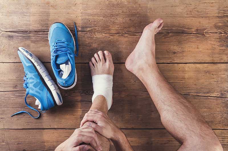
Injuries are frustrating, but whether you are a serious athlete or a weekend warrior, whether you play on your high school team or enjoy a pickup game at the YMCA, you will likely experience injury at some time. At Bluegrass Foot Centers we treat sports and fitness related injuries to the feet and lower extremities on a daily basis. Most commonly, we work with runners, cyclists, gymnasts, dancers, and soccer, football, baseball, basketball, and tennis players.
The most common injuries resulting from overuse are stress fractures, heel pain, Achilles tendinitis, ankle sprains, and Morton’s neuromas. While these conditions can be treated without casts or surgery, they do require close medical supervision. Very often, the treatment of sports injuries begin with an understanding of the underlying biomechanics that causes them. Dr. Block will perform a comprehensive biomechanical evaluation to determine how to best control these biomechanical forces, to allow for a quick recovery and to prevent further injury.
If left untreated or if exercise is resumed prematurely, the injury can be further aggravated. Our goal is to work together with you to minimize your downtime, if any, and get you healed as quickly as possibly. We will also examine your training habits and take measures to prevent injuries from reoccurring. And know this…we won’t make you stop your training unless it is absolutely necessary.
request an appointment